Sentinel Lymph Node Biopsy using ICG Fluorescence Imaging
A precise, minimally invasive technique for accurately staging cancer spread to lymph nodes — using indocyanine green (ICG) fluorescence imaging to identify and remove only the nodes most likely to contain disease, reducing the need for extensive lymph node removal.
- ICG Fluorescence-Guided Mapping
- Sentinel Node Identification
- Minimally Invasive Lymph Node Staging
- Breast, Gynaecologic & Other Solid Tumour Applications
- Reduced Lymphedema Risk
Overview
Accurate staging with less extensive surgery
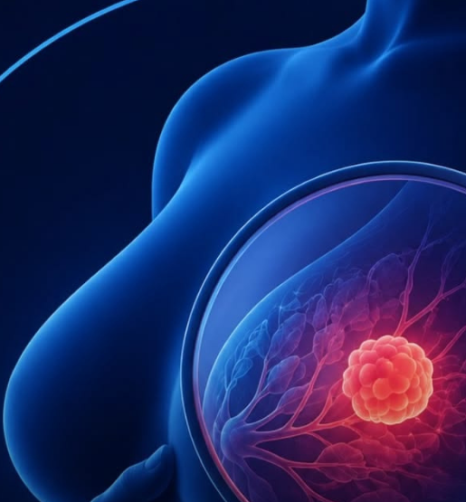
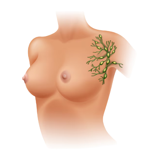
Knowing whether cancer has spread to nearby lymph nodes is one of the most important pieces of information in cancer staging — it shapes decisions about further treatment and prognosis. Traditionally, this has meant removing a larger number of lymph nodes to check them, which carries a real risk of complications such as lymphedema, numbness, and delayed healing. Sentinel lymph node biopsy offers a more targeted alternative, identifying the first, most likely node — or nodes — that cancer would spread to, and examining those specifically.
Indocyanine green (ICG) is a fluorescent dye injected near the tumour site, which travels through the lymphatic channels to the sentinel node. Using a specialised fluorescence imaging camera, the surgical team can visualise this pathway in real time, precisely identifying and removing the sentinel node or nodes for pathological examination — often during the same operation as the primary tumour surgery. This targeted approach provides highly accurate staging information while significantly reducing the extent of lymph node dissection required.
ICG fluorescence-guided sentinel node biopsy is used across a range of cancers, including breast cancer, endometrial and cervical cancer, and melanoma, and is incorporated into the surgical plan as determined by the multidisciplinary tumour board based on tumour type, location, and stage.
What We Offer
The ICG-Guided Sentinel Node Procedure
A precise, well-established technique integrated into your primary cancer surgery.
ICG Dye Injection
A small amount of indocyanine green dye is injected near the tumour site, timed to allow uptake into the lymphatic channels before surgery begins.
Fluorescence-Guided Lymphatic Mapping
Real-time visualisation of the dye as it travels through lymphatic channels, allowing the surgical team to trace the exact pathway to the sentinel node.
Sentinel Node Identification & Excision
Precise identification and removal of the first node, or nodes, in the lymphatic pathway most likely to contain cancer spread.
Intraoperative Pathological Assessment
Rapid examination of the removed sentinel node during surgery in select cases, helping guide further surgical decisions in real time.
Combined Sentinel Node Biopsy with Primary Tumour Surgery
Integration of sentinel node mapping into the same operation as tumour removal, avoiding the need for a separate procedure.
Application Across Breast, Gynaecologic & Skin Cancers
Use of ICG fluorescence mapping across multiple cancer types, tailored to the specific lymphatic drainage pattern of each tumour site.
Completion Lymph Node Dissection (When Indicated)
More extensive lymph node removal if the sentinel node is found to contain cancer, performed as a follow-on step where required.
Minimally Invasive & Robotic-Assisted Sentinel Node Mapping
ICG fluorescence mapping performed through laparoscopic or robotic-assisted approaches, combining precise node identification with a minimally invasive technique.
Bilateral & Multi-Site Nodal Mapping
Mapping and biopsy of sentinel nodes on both sides of the body or across multiple lymphatic basins, used in tumours with more than one possible drainage pathway.
Our Approach
The Sentinel Node Advantage
Accurate staging with a technique designed to minimise surgical impact.
A clear, supported path from first consultation through to long‑term follow‑up.
Precise, Real-Time Visualisation

Fluorescence imaging allows the surgical team to trace lymphatic pathways directly during surgery, improving accuracy over traditional mapping methods.
Reduced Extent of Surgery

Targeting only the sentinel node or nodes reduces the amount of lymph node tissue removed, lowering the risk of complications such as lymphedema.
Reliable Staging Information

Sentinel node status provides accurate information on cancer spread, directly guiding decisions about further treatment.
Seamless Integration with Primary Surgery

Sentinel node biopsy is performed as part of the same operation as tumour removal, avoiding additional procedures or delays.
Indocyanine green is a well-established dye used safely in surgery for decades. Your surgical team will discuss any individual considerations, such as allergy history, before your procedure.
Only if the sentinel node is found to contain cancer. If it’s clear, no further lymph node removal is typically needed, which reduces your risk of complications like lymphedema.
In most appropriately selected cases, yes — sentinel node biopsy provides accurate staging without the more extensive risks associated with full lymph node dissection.
In carefully selected early-stage, low-grade cases, uterus-preserving management may be an option. Your gynaecologic oncology team will assess your eligibility based on tumour characteristics.
Sentinel node biopsy is typically integrated into the same operation as your primary tumour surgery, adding relatively little additional time while providing valuable staging information.
Contact
Discuss Your Treatment Options
Every case is unique. Book a consultation to understand the safest and most personalised path forward for you.
Address
# 69, IOA Complex, Royapettah High Rd, Royapettah, Chennai, Tamil Nadu 600014
info@support.com
Our Number
+(0361) 234 567